


Surgeons face significant challenges when locating small lung nodules, particularly those under 8 millimeters that may indicate early-stage lung cancer. Traditional CT-guided methods require multiple scans and lengthy procedures, increasing the risk of complications. Dr. Wang Jiyong, head of thoracic surgery, highlights the limitations of conventional approaches, noting that even clear CT images do not allow for precise identification of tiny lesions.
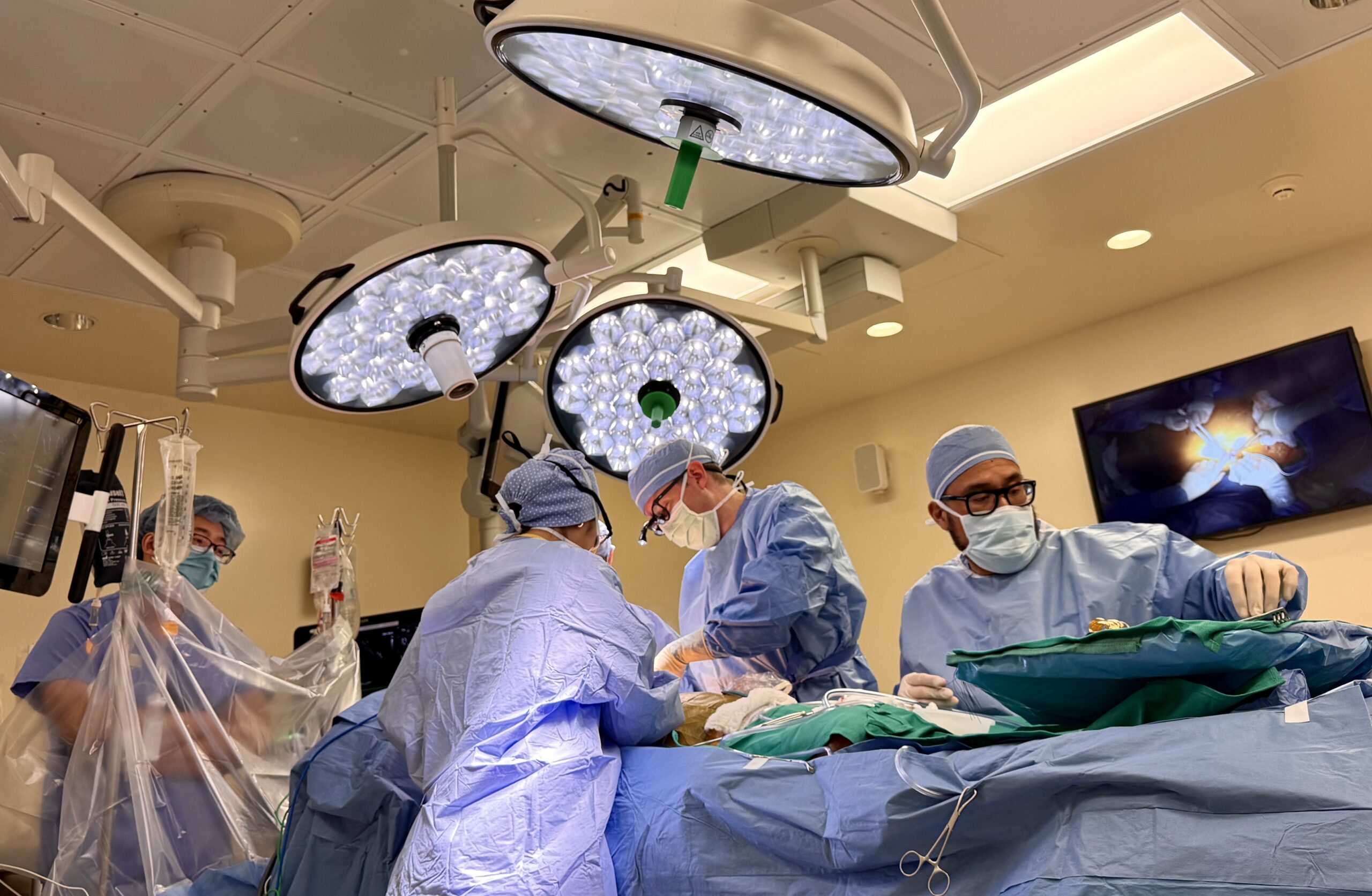
AI-driven surgical robots are transforming this landscape by utilizing 3D imaging to gather lesion data and create a spatial model of the lungs. This technology, likened to a high-precision GPS navigation system, enables pre-surgical planning of optimal puncture angles and real-time tracking of instruments during surgery. This advancement promises reduced trauma and shorter recovery times for early lung cancer patients.
As the detection rate of lung nodules rises with increased health screenings, Dr. Wang advises regular monitoring for nodules under 8 millimeters and vigilance for those above 5 millimeters. The evolution of AI robots from diagnostic tools to navigational aids is reshaping the surgical approach to lung nodules, making previously daunting tasks more manageable.
Editor's Note
The integration of AI in surgical procedures is revolutionizing the healthcare sector, particularly in thoracic surgery. This advancement not only enhances precision but also minimizes patient risks associated with traditional methods. As AI technology continues to evolve, its applications in real-time navigation during surgeries could set new standards in surgical practices and patient outcomes.

























Leave a comment