

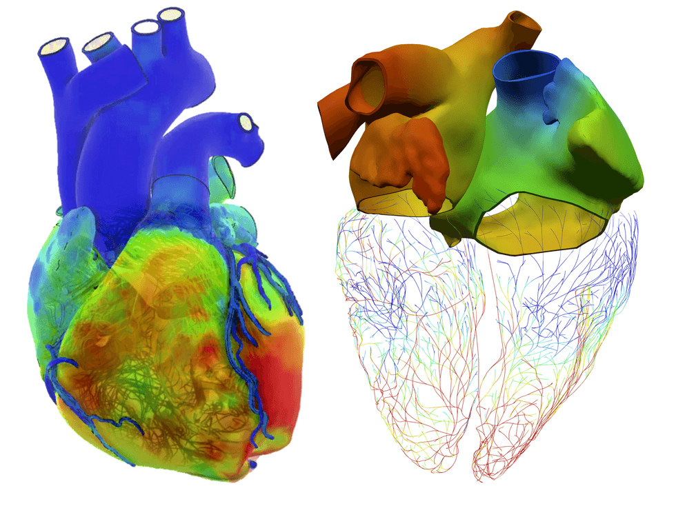
In May 2019, a cardiac surgeon at Boston Children’s Hospital successfully performed a complex heart surgery on a child with a severe congenital defect, utilizing advanced virtual twin technology. This innovative approach involved creating a detailed 3D model of the child's heart and vascular system from MRI and CT scans, allowing the surgical team to simulate various strategies and predict outcomes before the operation. The procedure was critical due to the unique nature of the child's heart condition, which had no established surgical manual.
The Living Heart Project, initiated in 2014, has since guided nearly 2,000 surgeries by employing virtual twin modeling, which combines engineering principles with medical expertise to enhance surgical precision and patient outcomes. This project, now involving over 150 organizations globally, aims to revolutionize medical treatment by providing a dynamic, predictive tool that can simulate the human body's responses.
The technology not only aids in surgical planning but also has the potential to streamline clinical trials. By creating virtual patient cohorts, researchers can test treatments more efficiently, reducing the time and costs associated with traditional trials. The FDA has recognized the significance of this approach, collaborating with the project to establish guidelines for in silico clinical trials, marking a significant shift in how medical innovations are developed and validated.
As virtual twins expand beyond cardiac applications to other organs, they promise to transform healthcare by enabling personalized medicine and fostering a deeper understanding of patient physiology, ultimately improving treatment outcomes and patient engagement in their health management.





























Leave a comment